Medial Branch Block for Diagnostic Back Pain Evaluation: What to Expect
A medial branch block is a short, image-guided diagnostic procedure used to identify whether the small facet joints in the spine are a source of a patient's back or neck pain. It is not a long-term treatment on its own; instead, the clinical team uses the response to a temporary anesthetic to decide whether a longer-acting procedure, such as radiofrequency ablation, is likely to help.
The facet joints are paired joints at the back of each spinal segment that guide and limit motion. The medial branch nerves are small sensory branches that carry pain signals from these joints. When providers suspect that a patient's axial back pain, the deep, aching pain that worsens with extension, twisting, or prolonged standing, is coming from the facet joints, a medial branch block helps confirm or rule out that hypothesis before any longer-term step is considered.
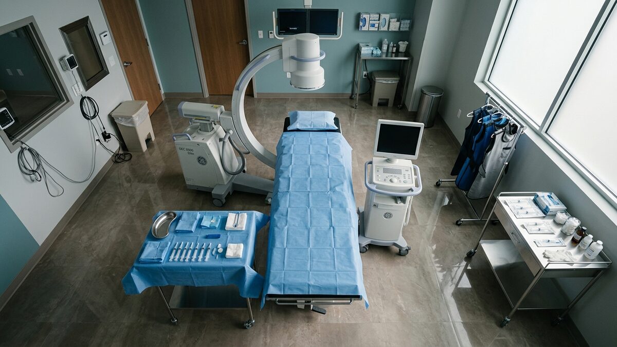
The procedure itself is brief. After check-in, patients are positioned face down on a fluoroscopy table, and a small area of skin over the targeted vertebral level is cleaned and numbed with a local anesthetic. Using live X-ray guidance, the provider directs a thin needle to the bony landmark where the medial branch nerve crosses the vertebra. A small volume of local anesthetic is then deposited adjacent to the nerve. Patients typically feel pressure during needle placement rather than sharp pain, and the contact portion of the procedure usually lasts only a few minutes per level.
The diagnostic value of a medial branch block comes from a defined observation window after the injection. Because the anesthetic used is short-acting, its effect is concentrated in the first several hours. Patients are usually asked to keep a simple pain diary during this window, rating their usual activities, sitting, standing, walking, bending, against their baseline pain. The clinical team is not looking for permanent relief from this injection. They are looking for a clear, time-limited reduction in the specific pain pattern the patient came in with.
How the clinical team interprets the response and what comes next
Interpretation is structured rather than impressionistic. A meaningful positive response is generally defined as a substantial reduction in the target pain during the expected window of anesthetic effect, with a return of pain afterward. Many practices use a two-block protocol, repeating the diagnostic injection on a separate day, to reduce the chance that a single positive result was driven by placebo response or by anesthetic spreading to a nearby structure. A consistent positive response across both blocks raises confidence that the facet joints are in fact the pain generator.
When the diagnostic response is consistent and positive, providers may discuss radiofrequency ablation, sometimes called facet rhizotomy, as a possible next step. That procedure uses heat to interrupt the same medial branch nerves for a longer period, often months. When the response is partial, ambiguous, or negative, the clinical team treats that as useful information too: it suggests the facet joints are unlikely to be the dominant source, and the workup shifts toward other potential contributors such as disc-related pain, sacroiliac joint dysfunction, or muscular and postural factors. In either case, the diagnostic block has done its job by narrowing the differential.
Preparation is straightforward. Patients are typically asked to arrive with a driver, to wear loose clothing that allows access to the back, and to confirm their current medication list with the scheduling team in advance. Anticoagulant and antiplatelet medications, certain supplements, and some diabetes medications may need adjustment before any image-guided injection; these decisions are individualized and coordinated between the patient's prescribing physician and the proceduralist. Patients who have had recent infections, fevers, or skin changes near the planned injection site should report this when confirming the appointment, since the procedure may need to be rescheduled.
After the injection, most patients are observed briefly and then discharged the same day. Mild soreness at the injection site is common for a day or two and is generally managed with ice and rest. The pain diary remains the most important part of the recovery period, and providers will often schedule a short follow-up to review it together. Driving and return to routine activity depend on individual circumstances and the type of sedation, if any, that was used, and patients receive specific instructions at discharge.
A medial branch block is best understood as a diagnostic tool rather than a treatment endpoint. Its value lies in producing clearer information about where pain is, and is not, originating, so that subsequent recommendations rest on observed response rather than assumption. Patients who respond well to a properly conducted diagnostic block, and who continue to have facet-pattern pain afterward, often have a clearer path forward in their care plan.
This article is informational and is not medical advice. Treatment options should always be made in consultation with a qualified physician.